The imaging and analysis team provides a wide range of tools, training, and expertise, with a primary focus on providing access to world-class advanced electron microscopy instrumentation for both materials characterization and biological imagingto the CNS community.
The Imaging & Analysis Suite is a satellite of the CNS (Center for Nanoscale Systems), a shared-use core facility. This Allston-SEC core provides researchers with access to SEM and TEM/STEM imaging, elemental analysis using EDS, and a suite of processing equipment including ultra-microtomes, sputter coater, polishing wheels, ion mills, and more to meet your sample preparation needs. The CNS is a member of the National Science Foundation’s National Nanotechnology Infrastructure Network (NNIN) initiative to create a national network of world-class facilities available to all researchers.
Onsite availability 9 am – 5 pm, M-F
Onboarding
To use CNS facilities, researchers must enroll as CNS users: https://cns1.rc.fas.harvard.edu/become-cns-user
A list of upcoming trainings can be found here: https://cns1.rc.fas.harvard.edu/training-sign-up/
Trainings may also be scheduled by emailing agraham@cns.fas.harvard.edu or Nwatson2@fas.harvard.edu
Fees Rates, billing policy, and other information can be found at https://cns1.rc.fas.harvard.edu/user-info/
About the team
Adam Graham supports the imaging and analysis team within CNS. He specializes in Cryo SEM, Cryo Sample preparation, high resolution TEM, Aberration corrected TEM, STEM and general sample pre/imaging needs. Adam provides training, application support, and assisted work as needed for CNS users. Before coming to CNS, Adam Supported the electron microscopy industry, performing sales, installations, training, applications support, and service to the global community.
Nicki Watson has been an electron microscopist for over 30 years. She specializes in biological electron microscopy imaging and sample prep and offers special assistance and training to researchers needing this technique. Before coming to CNS, Nicki managed the W.M.Keck Biological Imaging Facility at Whitehead Institute (1998-2019) . Nicki graduated from SDSU, spent two years working as a clinical electron microscopist at the VA Medical Center in La Jolla, California, followed by six years training with Drs. George Palade and Marilyn Farquhar at the University of California San Diego, as part of their electron microscopy core group.
Contact Email
Adam Graham agraham@cns.fas.harvard.edu
Nicki Watson Nwatson2@fas.harvard.edu
CNS
To learn more about the CNS in both Cambridge and Allston, visit CNS’s main page.
Equipment
Electron Microscopes:
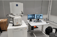
Zeiss Gemini 360 FE-SEM SEC
SEC-SEM-12
Imaging and Analysis
Make Zeiss
Model Gemini 360
LOCATION Allston SEC Imaging Suite LL2.301-03
Contact Adam Graham for training
The Gemini 360 Field Emission Scanning Electron Microscope (FESEM) enables high resolution surface examination and analysis. This SEM has a cross-over free Zeiss Gemini 1 column, and uses a low to moderate energy (0.02 to 30 keV) electron beam to image samples in high-vacuum with sub-nanometer resolution at 15 keV and 1.2 nm at 1 keV. Available detectors include integrated navigation camera In-lens SE; Everhart-Thornley detector; High Efficiency 6-segment Backscatter Detector; and Oxford Instruments Ultim Max EDS. The SEM is also equipped with a leica Cryo stage which enables imaging of frozen hydrated samples.
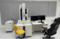
Hitachi 7800 TEM
SEC-TEM-12
Imaging and Analysis
Make Hitachi High Technologies
Model HT7800
LOCATION Allston SEC Imaging Suite LL2.301-05
Contact Nicki Watson for training
The HT7800 TEM is a 120 kV transmission electron microscope (TEM) with multiple lens configurations, including an unsurpassed high contrast optics design and is especially designed for easy operator use and fast sample exchange. The system provides high contrast imaging and is perfect for biological samples. The HT7800 TEM has a multi sample holder (can hold three at once) and can also be used for tomography data set acquisition. The breakthrough in advanced innovative design allows for highly efficient workflows and many specialized applications. It represents the cutting-edge solution for modern TEM analyses.
Click here to view this tool in the CNS virtual reality model (in the model, the tool is still in the Cambridge facility, but it has now been moved to the Allston SEC facility).
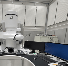
JEOL F200 TEM
SEC-TEM-13
Imaging and Analysis
MAKE JEOL
MODEL F200
LOCATION Allston SEC Imaging Suite LL2.301
Contact Adam Graham for training
The JEOL F200 is a field emission TEM/STEM. TEM Resolution spec is 1 angstrom at 200kv 1.6A for STEM. The system can work at both 200kv and 80kv It is equipped with a One View Camera, Bright field and HAADAF detector for STEM as well as a JEOL EDS detector with Oxford instruments Aztec software as the user interface. The F200 is also equipped with Serial EM to perform tomography with higher tilt limits when a High tilt holder is used. Standard holders are limited to lower tilt angles.
Sample prep tools:
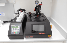
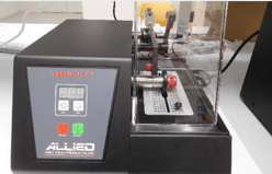
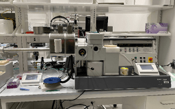
Allied Polisher (SEC)
SEC-HAR-059
Imaging and Analysis
MAKE Allied High Tech Products
MODEL System 8
LOCATION Allston SEC Imaging Suite LL2.301
Contact Adam Graham for training
The Allied MultiPrep System enables precise semi-automatic sample preparation of a wide range of materials for microscopic (optical, SEM, EBSD, FIB, TEM, AFM, etc.) evaluation. The AD-5™ Fluid Dispenser provides automatic, application of abrasive polishing suspensions or lubricants. Its function can be controlled through Allied’s MetPrep 3.
Allied Precision Low Speed Saw (SEC)
SEC-HAR-060
Imaging and Analysis
MAKE Allied High Tech Products
MODEL Tech Cut 4
LOCATION Allston SEC Imaging Suite LL2.301
Contact Adam Graham for training
The TechCut 4™ is a precision low-speed saw excellent for cutting smaller, delicate samples that cannot tolerate increased heat caused by high-speed sectioning. The pivoting cutting arm has adjustable weights to apply or counterbalance downward force to the sample during sectioning. Cutting fluid is drawn from the reservoir by the blade to cool the sample. With a 3″ to 6″ blade range, samples up to 2″ thick can be sectioned.
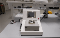
Critical Point Dryer Tousimis 931 GL (SEC)
SEC-CD-4
Imaging and Analysis
MAKE Tousimis
MODEL 931 GL 2.5
LOCATION Allston SEC Imaging Suite LL2.301
Contact Adam Graham or Nicki Watson for training
The Tousimis 931 GL critical point dryer has a 2.5″” chamber for larger samples/high volume drying and has a user-friendly touch screen user interface. It is equipped with state-of-the-art stasis software for drying gels and more challenging samples. The system is compatible with Biological and material samples.
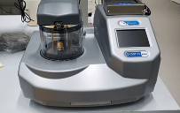
EMS 150T ES Sputter/Carbon Coater
SEC-HAR-061
Imaging and Analysis
MAKE Electron Microscopy Sciences
MODEL Q 150T ES Plus
LOCATION Allston SEC Imaging Suite LL2.301
Contact Adam Graham or Nicki Watson for training
This is a multi-purpose coater that can be used for sputter coating of conductive metals primarily for SEM. It can also be used as a high vacuum carbon coater using carbon rods and thread. The system can also work as a glow discharge unit and is fitted with a quartz monitor for measuring the thickness of deposition.
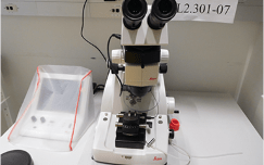
Leica UltraMicrotome SEC
SEC-HAR-062
Imaging and Analysis
MAKE Leica
LOCATION Allston SEC Imaging Suite LL2.301-07
Contact Nicki Watson for training
This dedicated room temperature microtome is designed to cut sections of embedded material for imaging in a TEM.
Leica Cryo-UltraMicrotome SEC
SEC-HAR-063
Imaging and Analysis
MAKE Leica
LOCATION Allston SEC Imaging Suite LL2.301-07
Contact Nicki Watson for training
This Ultra Microtome is a dedicated Cryo system for cutting sections of frozen material to be imaged in a cryo TEM.
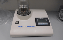
Pelco Easyglow
SEC-BIO-17
Imaging and Analysis
MAKE Pelco
LOCATION Allston SEC Imaging Suite LL2.301-07
Contact Nicki Watson for training
For Glow discharge of your EM grids and samples
Vitrobot
SEC-HAR-064
Imaging and Analysis
MAKE Unknown
LOCATION Allston SEC Imaging Suite LL2.301-07
Contact Nicki Watson for training
The Vitrobot is used for freezing your TEM sample using Liquid Ethane to preserve your samples under Cryogenic conditions forming Vitrified ICE.
Bal-tec Cryo Preparation
SEC-HAR-065
Imaging and Analysis
MAKE Bal-Tec AG
MODEL MED020
LOCATION Allston SEC Imaging Suite LL2.301
Contact Adam Graham for training
The MED 020 with freeze fracture chamber is used to prep your frozen hydrated sample for imaging in the FESEM.